Treeinbuds
TreeinBud sign Lungs
Usually somewhat nodular in appearance, the tree-in-bud pattern is generally most pronounced in the lung periphery and associated with abnormalities of the larger airways. Normal lobular bronchioles (≤ 1 mm in diameter) cannot be seen on CT scans, which can only show bronchi more than 2 mm in diameter. However, diseased bronchioles can be seen.

Tree in bud sign Radiology Case
Radiographic features. Tree-in-bud sign is not generally visible on plain radiographs 2 . It is usually visible on standard CT, however, it is best seen on HRCT chest. Typically the centrilobular nodules are 2-4 mm in diameter and peripheral, within 5 mm of the pleural surface. The connection to opacified or thickened branching structures.
Treeinbud sign radRounds Radiology Network
The tree-in-bud sign is a nonspecific imaging finding that implies impaction within bronchioles, the smallest airway passages in the lung. The differential for this finding includes malignant and inflammatory etiologies, either infectious or sterile. This includes fungal infections, mycobacterial infections such as tuberculosis or mycobacterium.

TreeInBud Pattern AJR
The 'tree-in-bud' sign is an unspecific sign of bronchiolar and alveolar pathology. Many of the more common differential diagnoses, particularly infectious causes, can be determined by analyzing the features of HRCT in conjunction with the patient history, the clinical presentation, and a microbiological analysis of the BAL.

Treeinbud sign Plugged small airways
The tree-in-bud sign indicates bronchiolar luminal impaction with mucus, pus, or fluid, causing normally invisible peripheral airways to become visible [80]. It is not specific for a single disease entity, but is a direct sign of various diseases of the peripheral airways and an indirect sign of bronchiolar diseases, such as air trapping or sub.

Treeinbud sign and bronchiectasis Radiology Case
The tree-in-bud sign is a nonspecific imaging finding that implies impaction within bronchioles, the smallest airway passages in the lung. The differential for this finding includes malignant and inflammatory etiologies, either infectious or sterile. This includes fungal infections, mycobacterial infections such as tuberculosis or mycobacterium.

Treeinbud sign and bronchiectasis Radiology Case
Journal of Thoracic Imaging: March 2012 - Volume 27 - Issue 2 - p W27. doi: 10.1097/RTI.0b013e31824643ae. Buy.

CT scan showing cavitary lesion in the right lung and treeinbud sign
Tree-in-bud: summary. Tree-in-bud opacities are seen on chest CT. They are small branching and nodular opacities which indicate disease of the small airways or arteries. They are most commonly seen with infections that involve the airways but have many causes. The treatment will depend on the underlying cause.

tree in bud opacities seen in Lettie Boisvert
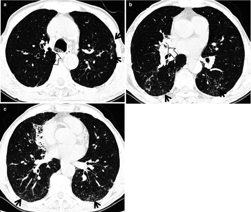
Case Discussion. The case shows left greater than right, predominantly basilar mixed consolidative airspace and patchy ground glass opacity intermixed with tree-in-bud type nodularity. Differential diagnosis: infectious bronchiolitis : MAC, bacterial and viruses. follicular bronchiolitis. panbronchiolitis.

tree in bud opacities in lungs Silvana Schilling
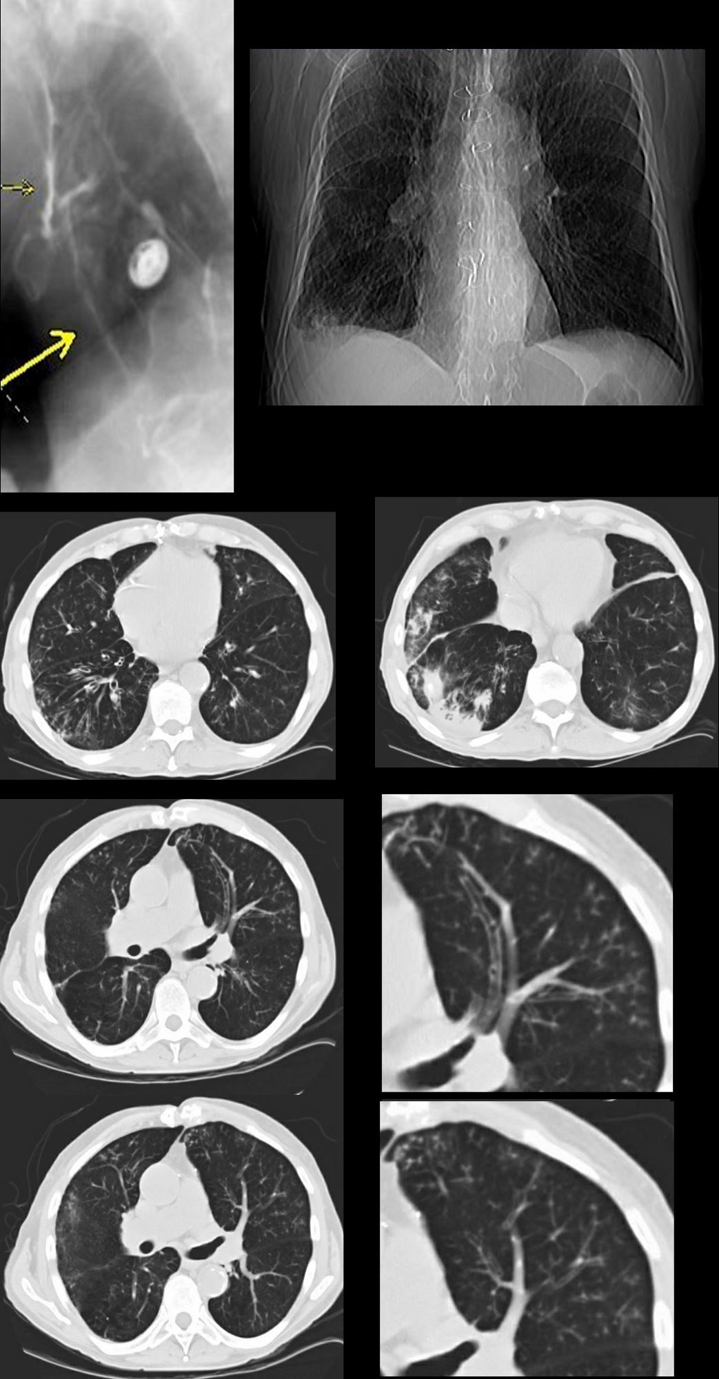
Results of the analysis of HRCT findings Tree-in-bud sign (TIB) Among the 200 cases of MPP, 174 cases showed the tree-in-bud sign (TIB) (Fig. 1, red arrow), accounting for 87%, compared with 62%.
TreeinBud Sign Radiology Key
Abstract. Tree-in-bud sign refers to the condition in which small centrilobular nodules less than 10 mm in diameter are associated with centrilobular branching nodular structures [1] (Fig. 9.1). The small nodules represent lesions involving the small airways. However, vascular lesions involving the arterioles and capillaries may simulate the.

TreeInBud Pattern AJR
In intralobular distribution of nodules, the "tree-in-bud" sign can be seen (↑) which is characterized as the presence of small well-defined foci at the ends of linear intralobular structures.This sign almost always indicates the presence of dilated and filled with some substrate (mucus, detritus, pus, cellular elements) intralobular bronchioles.

TreeInBud Pattern AJR
The 'tree-in-bud' sign is a common finding in HRCT scans. The list of the most frequent differential diagnoses for 'tree-in-bud' sign includes infections with Mycobacterium tuberculosis, nontuberculous mycobacteria, and other bacterial, fungal, or viral pathogens. Other causes could be immunological, congenital, and idiopathic disorders as well.

Pin on THORAX
Case Discussion. Chest x-ray in a 60 year old patient of Asian extraction demonstrates faint reticulonodular opacities. CT confims numerous centrilobular nodules with opacified distal bronchioles ( tree-in-bud sign) and bronchiectasis. These findings most likely represents pulmonary TB or MAC despite negative induced sputum specimens.

Tree in Bud Sign The TreeinBud sign represents Endobronchial
The tree-in-bud pattern is commonly seen at thin-section computed tomography (CT) of the lungs. It consists of small centrilobular nodules of soft-tissue attenuation connected to multiple branching linear structures of similar caliber that originate from a single stalk. Originally reported in cases of endobronchial spread of Mycobacterium tuberculosis, this pattern is now recognized as a CT.

tree in bud opacities causes Clothed With Authority Online Diary
Tree-in-bud sign. The tree-in-bud pattern represents bronchiolar luminal impaction with mucus, pus or fluid, which demarcates the normally invisible branching course of the distal peripheral pathways, thus resembling a tree branch studded with buds on High-Resolution CT of the lungs (Figs. 1, 2) [ 1 ]. This sign corresponds to small (2-4 mm.